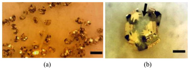
Fig. 13.

Bright-field optical microscopy images of deployed μ-grippers on a fresh pig stomach tissue. (a) 10 min after the deployment. The scale bar = 1 mm. (b) Close-up image showing a μ-gripper latching onto a piece of tissue. Arrow points out semitransparent tissue and the scale bar represent 100 μm.
