
FIG 6.
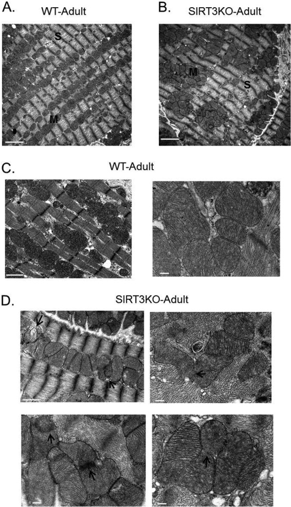
Transmission electron micrographs showing mitochondrial morphology in SIRT3KO hearts. Electron micrographs showing mitochondrial morphology and arrangement in adult wild-type (WT) (A and C) and SIRT3KO hearts (B and D). SIRT3KO micrographs show mitochondria clustered in islands (B). Arrows in SIRT3KO micrographs (D) indicate defective mitochondrial inner membrane fusion. M, mitochondria; S, sarcomeres. Scale bars: 2 μm (A and B); 1 μm (C [left panel] and D [left top panel]); and 200 nm (C [right panel] and D [bottom and right top panels]).