
Figure 8. Quantitative Western blot analysis of the levels of PGC-1α, TFAM, NRF-1, and oxidized proteins in DRG from PGC-1α (+/+) and PGC-1α (−/−) mice with and without diabetes.
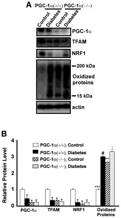
A. Protein extracts (25 μg of protein/lane) from DRG of PGC-1α (+/+) non-diabetic control and 1month diabetic mice, and PGC-1α (−/−) non-diabetic control and 1 month diabetic mice were subjected to one-dimensional gel electrophoresis followed by immunoblotting with antibodies against PGC-1α, TFAM, NRF-1, DNP, and actin. B. The relative level of each protein was normalized to the actin level in the corresponding samples. The bar graph shows the results (mean ± SEM) from five PGC-1α (+/+) non-diabetic control and diabetic mice, and PGC-1α (−/−) non-diabetic control and diabetic mice. *P <0.05 for a decrease in the levels of PGC-1α, TFAM and NRF-1 in PGC-1α (+/+) diabetic mice, or PGC-1α (−/−) diabetic and control mice compared to PGC-1α (+/+) non-diabetic control mice. ***P <0.001 for an increase in oxidized proteins in PGC-1α (+/+) diabetic compared to PGC-1α non-diabetic (+/+) mice, ⊥ P <0.01 for PGC-1α (−/−) diabetic compared to PGC-1α (+/+) diabetic mice, # P <0.05, PGC-1α (+/+) diabetic compared to PGC-1α (−/−) non-diabetic mice, **P <0.01 for an increase in oxidized proteins in PGC-1α (−/−) diabetic mice compared to PGC-1α (−/−) non-diabetic mice.