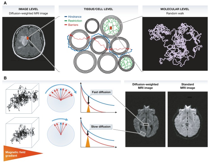
Figure 1.
(A) Contrast and signal levels in the diffusion-weighted image (left) reflect water diffusion behavior (random walk) (right). Diffusion behavior is modulated by tissue structure at the cellular level (middle): For instance, diffusion can be restricted within cells, water may escape when cell membranes are permeable and might then experience a tortuous pathway in the extracellular space (hindrance). (B) In the presence of a magnetic field gradient (variation of the magnetic field along one spatial direction), magnetized water molecule hydrogen atoms are dephased. The amount of dephasing is directly related to the diffusion distance (a few micrometers) covered by water molecules during measurement (a few tens of milliseconds). Given the great many water molecules experiencing individual random walk displacements, the overall effect of this dephasing is an interference, which reduces MRI signal amplitudes. In areas with fast water diffusion (e.g. within ventricules), the signal is deeply reduced, while in areas of slow water diffusion (e.g. white matter bundles), the signal is only slightly reduced. This differential effect results in a contrast in the diffusion-weighted MRI images, which is not visible in standard MRI images.

