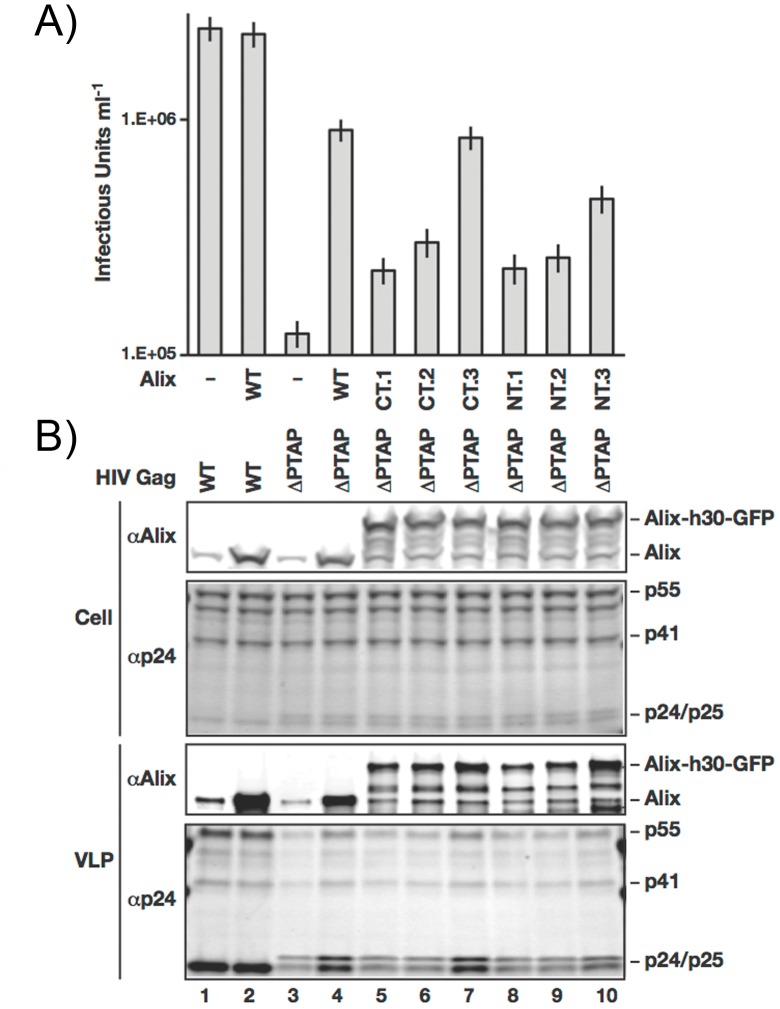
Figure 2. Characterization of the ALIX fusion proteins with eGFP.
A) Cells expressing ALIX fusions (lanes 5–10), wt ALIX (lanes 2 and 4) or control (lanes 1 and 3) where infected with WT (Lanes 1 and 2) as well as ΔPTAP HIV-1 RΔ8.2 (Lanes 3–10). Assembled virions were harvested 16 hrs post infection and viral titers were measured using FACS to detect eGFP expression from the packaged pLOX-GFP vector in transduced HeLa cells. B) Western blot analysis of cells and collected virions from the same experiment presented in A.

