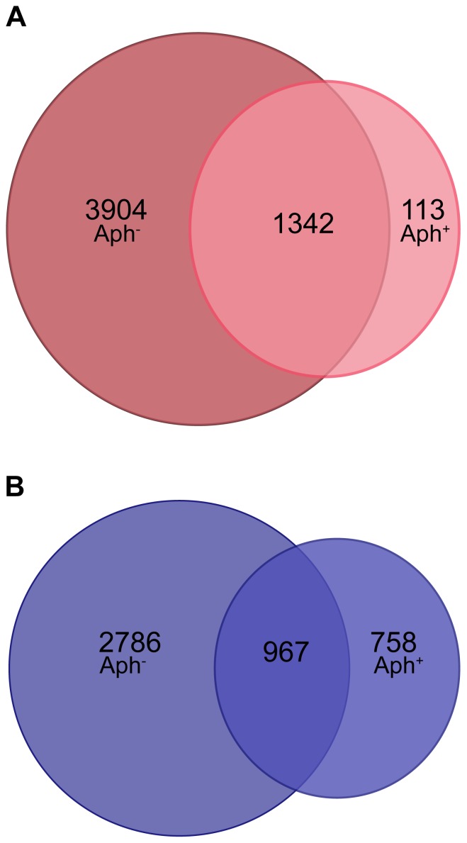
Figure 3. Venn diagram of transcripts elevated ≥2-fold in embryos treated or not treated with aphidicolin.
Pronuclear oocytes were cultured in the presence of aphidicholin (Aph) from 6 to 96 hours post activation (hpa), and compared with sibling embryos that were not treated so as to appreciate how many mRNAs are upregulated regardless of the cell cycle progression. (A) Venn diagram showing mRNAs that accumulate after SCNT when the first round of embryonic DNA replication is suppressed (Aph+) in comparison to mRNAs that accumulate when the embryo can cycle normally (Aph−). (B) Venn diagram showing mRNAs that accumulate after fertilization when the first round of embryonic DNA replication is suppressed (Aph+) in comparison to mRNAs that accumulate when the embryo can cycle normally (Aph−). In both SCNT and fertilization, mRNAs were considered whose abundance increased at least 2-fold compared to MII oocytes.

