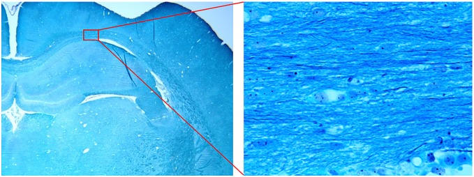
Figure 7. Typical microscopic images with 2x (left) and 100x (right) magnifications taken from ROI#5 of one of our cases.

Nuclei are colorless; myelin is blue and axons are black in the stained sections.

Nuclei are colorless; myelin is blue and axons are black in the stained sections.