
Figure 3.
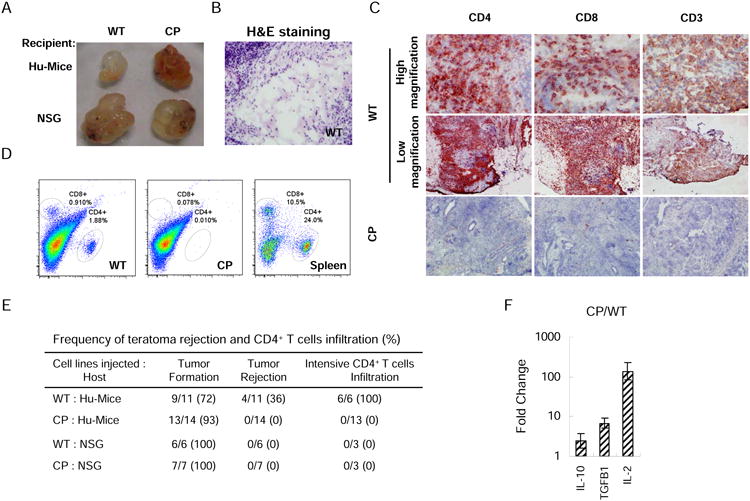
Expression of PD-L1 and CTLA4-Ig protects CP hESC-derived teratomas from allogeneic immune rejection. (A) Images of teratomas derived from WT and CP hESCs formed in Hu-mice and NSG mice. Mice were subcutaneously injected with WT hESCs and CP hESCs around the left and right hindlegs, respectively. Six-to-eight weeks after implantation, the mice were euthanized and teratomas examined. Representative images are shown. (B) Extensive tissue necrosis was detected in the teratomas formed by WT hESCs in Hu-mice. Significant T cell infiltration was detected in the teratomas formed by WT hESCs but not those formed by CP hESCs in Hu-mice as shown by immunohistochemistry (C) and flow cytometry (D). (E) Summary of teratoma formation, immune rejection and CD4+ T cell infiltration. Teratomas with apparent regressing phenotype or containing only liquid-filled cysts without cell mass were classified as rejection. (F) Relative mRNA levels of IL-10, TGFβ1 and IL-2 in T cells isolated from CP hESC- and WT hESC-derived teratomas formed in the same Hu-mouse were determined by real-time PCR. Mean values are presented with SD (N=3). See also Figure S2 and S3.