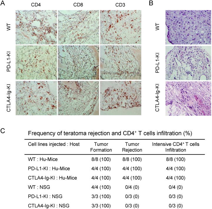
Figure 6.

Expression of PD-L1 or CTLA4-Ig alone cannot protect the derivatives of hESCs from allogeneic immune rejection. (A) Extensive T cell infiltration was detected in the teratomas formed by WT hESCs, PD-L1-KI-hESCs and CTLA4-Ig-KI-hESCs in Hu-mice. T cells were identified by anti-CD4, anti-CD8 and anti-CD3 antibodies. (B) Extensive necrosis was detected in the teratomas derived from WT hESCs, PD-L1-KI-hESCs and CTLA4-Ig-KI-hESCs in allogeneic Hu-mice, as revealed by hematoxylin and eosin staining. (C) The frequency of CD4+ T cell infiltration and teratoma rejection in Hu-mice transplanted with parental and various knock-in hESCs. See also Figure S5.
