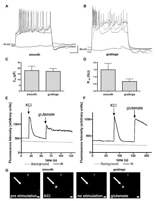
Figure 5. Action potentials of iNs generated on smooth substrates and 5 μm gratings.
(A) A representative trace of a train of action potentials in response to step depolarizing current injection of a synapsin-RFP+ iN on smooth substrate. (B) A train of action potentials in synapsin-RFP+ iN on 5 μm gratings in response to depolarizing current injection. (C) No difference was observed in membrane capacitance between iNs on smooth or grating substrates. (D) The input resistance of iNs on 5 μm gratings was slightly lower compared to iNs on smooth substrates. (E) A representative trace of fluorescent intensity measurements in iN on smooth and (F) on grating substrates in response of KCl and glutamate stimulation. (G) Image sequence of a representative iN (arrow) before stimulation, stimulated with KCl, between stimulations and stimulated with glutamate. Warm colors white, yellow and red represent high fluorescent intensities. Scale bar 10 μm.

