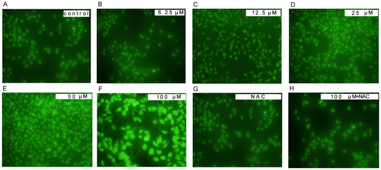
Figure 3. MEHP induced reactive oxygen species (ROS) generation in HUVEC cells.
In MEHP treatment group, the HUVEC cells were treated with 0 µM (A), 6.25 µM (B), 12.5 µM (C), 25 µM (D), 50 µM (E) and 100 µM (F) MEHP for 24 hours. In NAC+MEHP treatment group, the HUVEC cells were pretreated for 1 hour before the MEHP treatment and then treated with 0 µM (G) and 100 µM (H) MEHP for 24 hours. After cultured with 10 mM 2,7-dichlorofluoroscein diacetate (DCFH-DA), the HUVEC cells in both groups were photographed by a fluorescence microscope (400x). Data was collected from three independent experiments.

