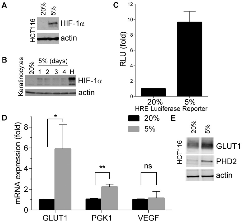
Figure 1. Physiological oxygen tensions induce HIF-1α expression and activity.
(A) Western blot showing the protein levels of HIF-1α in HCT116. Cells were cultured at 20% or 5% O2 for 72 hours. (B) Western blot showing the protein levels of HIF-1α in normal human keratinocytes cultured for 1 to 4 days at 5% O2 or treated 500 µM of chemical hypoximimetic CoCl2 for 16 hours (H). (C) Luciferase assay showing HIF-1α in HCT116 cells. HCT116 were transfected with a PGK-1 luciferase reporter plasmid and a β-galactosidase control plasmid and then cultured for 48 hours at 5% O2. β-galactosidase activity was used to normalize luciferase activity. Luciferase activity is expressed as a ratio to 20% O2 levels. Results show mean values of 3 independent experiments and error bars represent standard deviation (D) qRT-PCR showing mRNA levels of Glut-1, PGK-1 and VEGF in HCT116 cells cultured at 20% and 5% O2 for 24 hours. Results show mean values of 3 independent experiments and error bars represent standard deviation. P values (unpaired t-tests): 0.03 (*), 0.001 (**), 0.7 (ns). (E) Western blot of lysates of HCT116 cultured at 20% or 5% O2 for 72 hours, showing expression of Glut-1 and PHD2.

