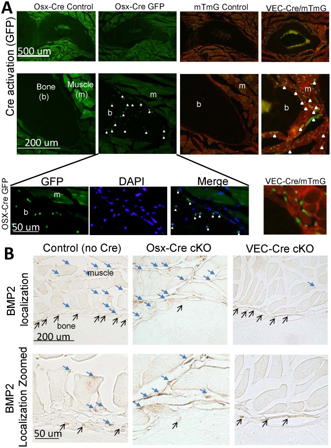
Figure 1. Deletion of BMP2 was evaluated in osteogenic cells (Osx-Cre) and vascular cells (VEC-Cre).
(A) First, Cre activation was verified by examination of cortical bone (ulna) and muscle from transverse cross-sections through the mid-forelimb. In control samples the bone is completely black. The Osx-Cre mouse has a GFP::Cre fusion protein which demonstrated the current expression of Cre in some osteocytes within the bone and osteoblasts lining the bone surface (open white arrowheads). The VEC-Cre mouse was crossed with mTmG reporter mouse and demonstrated Cre activation within muscle and periosteum compartments surrounding the bone (filled white arrowheads). (B) Confirmation of the BMP2 protein deletion was done using immunohistochemistry. In the control sample BMP2 expression was seen in the muscle (blue filled arrows) and bone-lining cells (thin black arrows). Using the Osx-Cre BMP2 expression was absent in the bone lining cells, but abundantly expressed throughout the muscle compartment (blue filled arrows). In contrast, using the VEC-Cre expression of BMP2 was limited to osteoblasts lining the bone surface (thin black arrows). Images are representative of 3–5 sections from 3–6 animals/group.

