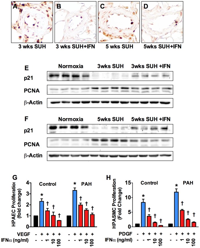
Figure 6. IFNα inhibits pulmonary vascular cell proliferation.
(A–D) Representative 40x images of lung sections from 3 week SUH rat, 3 week SUH rat + IFNα, 5 weeks SUH rat, and 5 week SUH rats+ IFNα stained for PCNA (brown) as an indicator of proliferating cells. WB analysis for PCNA and p21 in whole lung lysates from (E) 3-week SUH rats or (F) 5-week SUH rats with or without IFNα (n = 4 animals per group). (G) Control or IPAH HPASMC were serum starved 24 h and then stimulated with PDGF (10 ng/ml) with or without increasing IFNα for 24 hours. (H) Control or IPAH HPAEC were serum starved overnight and then stimulated with VEGF (50 ng/ml) with or without increasing IFNα for 24 hours. Proliferation was assessed by measuring [H3]-thymidine incorporation. Analysis of variance *P<0.05.

