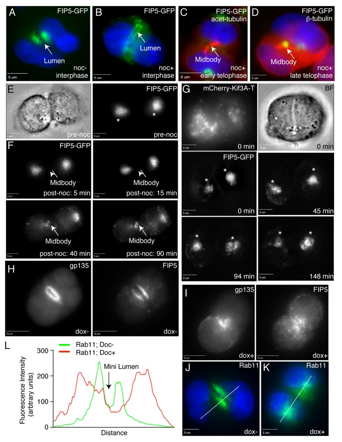
Figure 3. Kinesin-2 transports FIP5-endosomes along central spindle microtubules to the AMIS. (A–D) MDCK cells expressing FIP5-GFP were embedded into Matrigel and grown for 24 h. Cells were then incubated for 30 min in the presence (B–D) or absence (A) of 1 μM nocodazole, fixed and stained for either β-tubulin (D) or acetylated tubulin (C). Arrows point to either the lumen (A–B) or midbody (C–D). Scale bars: 5 μm. (E–F) MDCK cells expressing FIP5-GFP were embedded into Matrigel and plated on glass-bottom dishes. After incubation for 24 h, cells in early telophase (E–F) were chosen for time-lapse analysis. To depolymerize microtubules, 10 μM of nocodazole was then added and cells were imaged every five minutes for 90 min at 37 °C. Panels in (F) show selected images from time-lapse series. Arrow points to the midbody location. Scale bars: 5 μm. (G) MDCK cells transiently co-transfected with FIP5-GFP and mCherry-Kif3A-T were grown in 3D cultures for 24 h. Dividing cells were then analyzed by time-lapse microscopy. Asterisks mark FIP5 associated with centrosomes. Scale bars: 5 μm. (H–L) MDCK-shKif3A#1 cells were pre-incubated with or without doxycycline for 3 d and then grown in 3D cultures for 24 h in the presence (I and K) or absence (H and J) of doxycycline. Cells were then fixed and stained with anti-gp135 (H and I), anti-FIP5 (H and I) or anti-Rab11 (J and K) antibodies. Scale bars: 10 μm. Figure in (L) shows line-scan quantifications of Rab11 distributions (marked by lines in J and K).

An official website of the United States government
Here's how you know
Official websites use .gov
A
.gov website belongs to an official
government organization in the United States.
Secure .gov websites use HTTPS
A lock (
) or https:// means you've safely
connected to the .gov website. Share sensitive
information only on official, secure websites.
