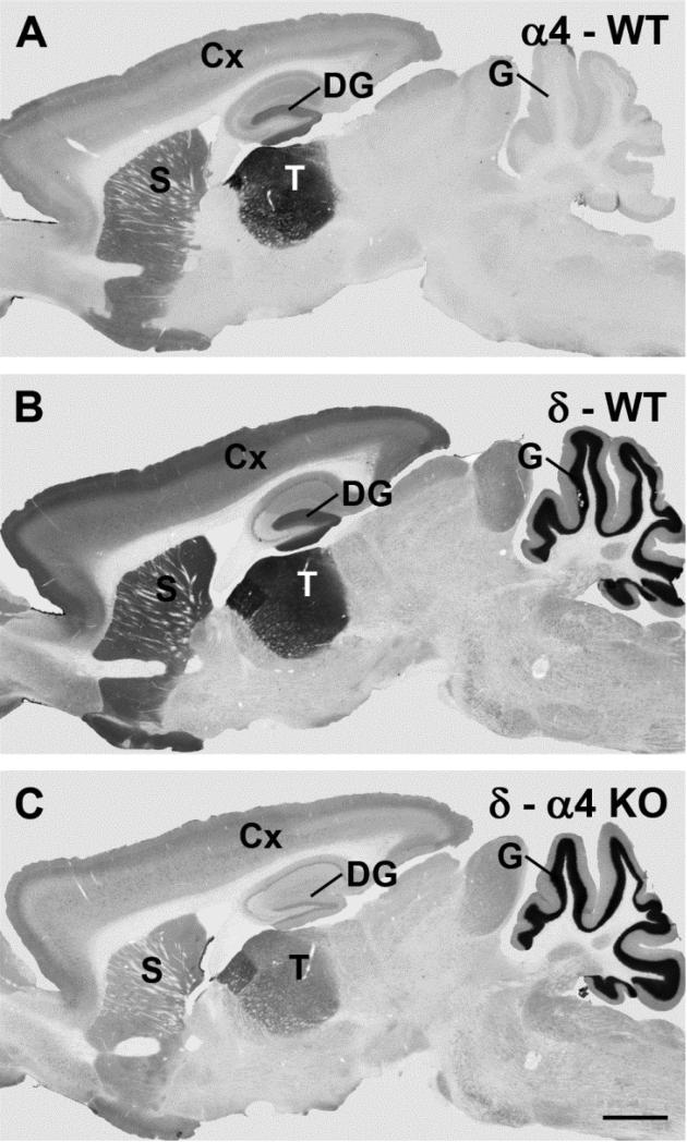
Fig. 1.
Comparisons of immunolabeling for the α4 and δ subunits of the GABAA receptor (GABAAR) in wild-type (WT) and α4 knockout (KO) mice in sagittal brain sections. a In an α4 KO mice, specific α4 subunit labeling is absent throughout the brain (labeled regions are identified below). b In a WT mouse, the α4 subunit is moderately to strongly labeled in specific forebrain regions, with the highest expression in the thalamus (T), molecular layer of the dentate gyrus (DG), striatum (S) and outer layers of the cerebral cortex (Cx). Virtually no specific α4 subunit labeling is evident in the granule cell layer (G) of the cerebellum. c In a WT mouse, δ subunit labeling closely parallels the pattern of α4 labeling in the forebrain, but is also present at high levels in the granule cell layer of the cerebellum. d In an α4 KO, δ subunit labeling is substantially reduced in all forebrain regions in which the α4 subunit is normally expressed. In contrast, no decrease in δ labeling is evident in the cerebellum which lacks α4 labeling in WT mice (see Panel B). Scale bar = 1 mm for A-D. (Comparisons of α4 subunit labeling in WT and α4 KO mice were originally described in Chandra et al., 2006).

