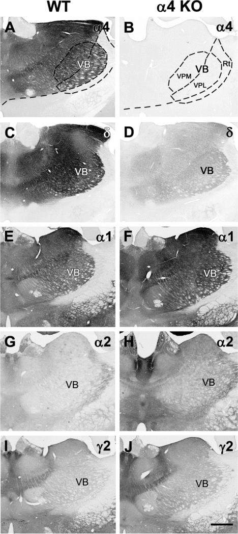
Fig. 2.
Comparisons of immunolabeling of GABAAR subunits (α4, δ, α1, α2, γ2) in coronal sections of the thalamus in WT and α4 KO mice. a,b In a WT mouse, strong α4 subunit labeling is present in many thalamic nuclei, including the ventrobasal (VB) complex which includes the ventral posterior medial (VPM) and ventral posterior lateral (VPL) nuclei, but is virtually absent from the nucleus reticularis (Rt). In the α4 KO, no specific labeling is present in the VB nucleus or other thalamic regions where strong α4 subunit labeling is normally present. c,d In a WT mouse, strong δ subunit labeling is evident in the thalamus and closely resembles the pattern of α4 labeling in WT mice. The δ subunit labeling is substantially reduced throughout these regions in the α4 KO. e,f In a WT mouse, α1 labeling is present at moderate levels in much of the thalamus including the VB nuclei and is increased within the same regions in the α4 KO. g,h In a WT mouse, α2 subunit labeling is low throughout much of the thalamus, including the VB nuclei, with the strongest labeling in the Rt and midline nuclei. Labeling is increased slightly throughout the thalamus in the α4 KO. i,j In a WT mouse, γ2 labeling is relatively low in much of the thalamus, including the VB nuclei. Some increases in γ2 labeling are evident in the α4 KO. Scale bar = 500 μm for A-J.

