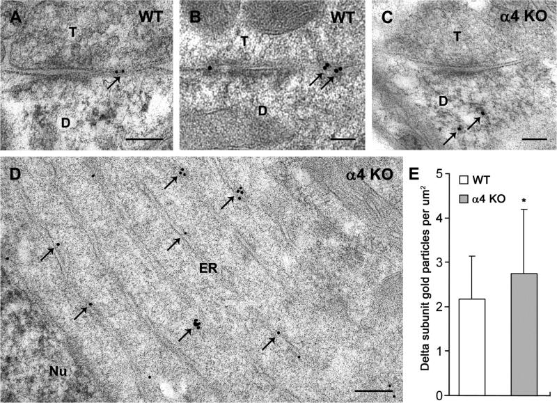
Fig. 6.
Comparison of immunogold labeling of the δ subunit in electron micrographs of the ventrobasal (VB) nucleus in WT and α4 KO mice. a,b In WT mice, δ subunit labeling is located predominantly at perisynaptic and extrasynaptic locations (arrows) near synaptic contacts between axon terminals (T) and postsynaptic dendrites (D). c In an α4 KO mouse, labeling along the plasma membranes near synaptic contacts appears reduced, and immunogold particles are evident within the cytoplasm (arrows). d In an α4 KO mouse, immunogold labeling for the δ subunit (arrows) is present within the cytoplasm of the cell body, and clusters of immunogold particles are evident near stacks of endoplasmic reticulum (ER), adjacent to the neuronal nucleus (Nu). e Comparisons of immunogold labeling in neuronal cell bodies of the VB nucleus indicate a higher concentration of immunogold particles in the cytoplasm in α4 KO mice than in WT mice (mean ± s.e.m., p < 0.05). Scale bars = 0.1 μm for A-C; 0.2 μm for D.

