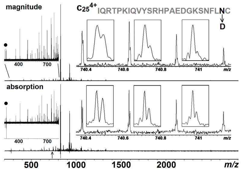
Figure 5.
ECD spectrum (MS3) of the b 9+63 fragment from β2 microglobulin in both magnitude- and absorption-mode. The inserts on the right are zoom in of the c4+25 fragment (labelled with an arrow in the bottom) with its sequence and the deamidation site highlighted in black. The inserts on the left are zooms of m/z 250-820 to show the entire spectrum is properly phased, and the peaks labelled with a dot are 3rd harmonic peaks of the peaks in m/z ~823.

