Figure 3.

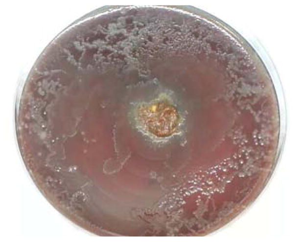
Strongyloides stercoralis. Stool inoculated in the center of a blood agar plate shows stool bacterial colonies in numerous serpiginous tracts, consistent with the presence of multiple live, motile larvae.

Strongyloides stercoralis. Stool inoculated in the center of a blood agar plate shows stool bacterial colonies in numerous serpiginous tracts, consistent with the presence of multiple live, motile larvae.