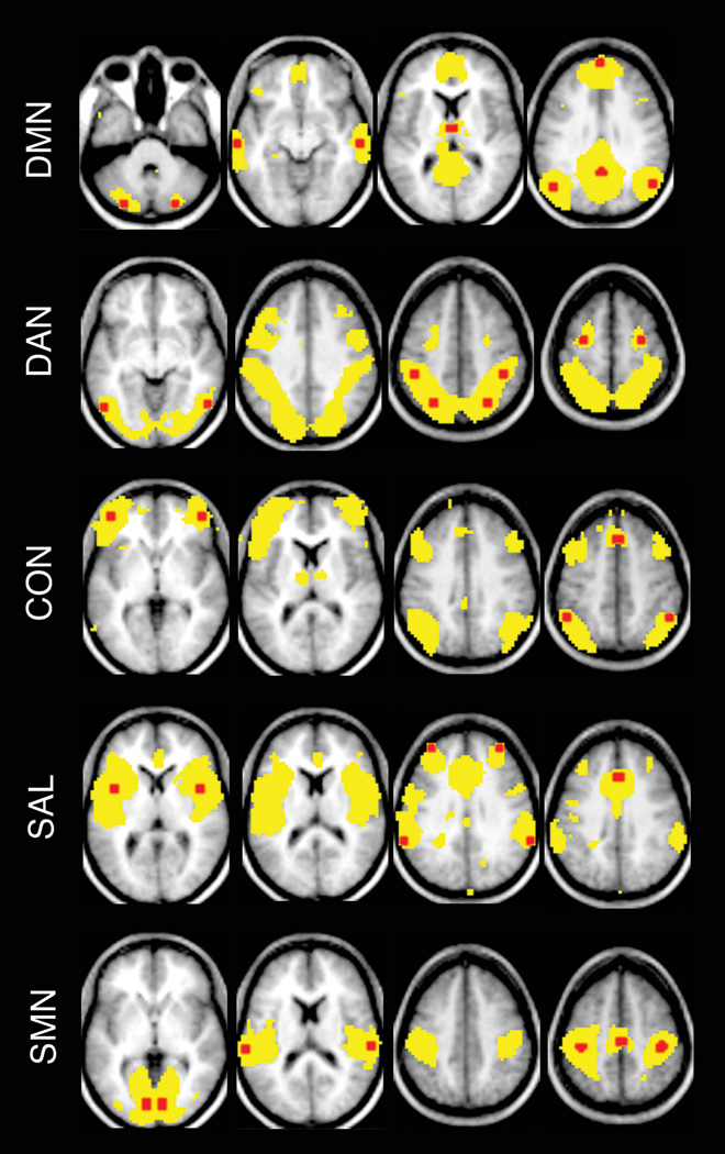
Figure 1.

Topographies of resting state networks (RSNs) and locations of seed regions.
Using a representative seed for each RSN, correlation maps were created for all AD participants. Group average maps thresholded at z(r)>0.1 are shown using yellow. All a priori seed regions (6-mm spheres) (red) from a RSN are overlaid on the group average map. Atlas coordinates for each seed region are provided in Supplemental Table S1. In particular, the posterior cingulate cortex was used as seed region for default mode network (DMN); the left posterior intraparietal sulcus for dorsal attention network (DAN); the left anterior prefrontal cortex for control network (CON), the left insular cortex for salience network (SAL), and the left primary visual cortex, the left primary auditory cortex and the left motor cortex for the sensory-motor network (SMN).
