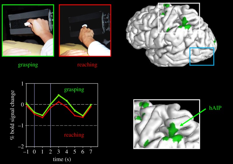
Figure 1.
Neuroimaging of visually guided grasping. The two panels in the upper left show the ‘grasparatus’ that James et al. [17] used to present graspable objects to participants in the scanner. Different rear-illuminated target objects could be presented from trial to trial on the grasparatus by rotating the pneumatically driven eight-sided drum. Participants were instructed either to grasp the object or to simply reach out and touch it with the back of the hand. Note that the experiment would normally be run in the dark with only the illuminated target object visible. The graph at the bottom left side shows the time course of activations in hAIP when a healthy participant reached out and grasped an object (green) versus when she reached out and simply touched the object with the back of her hand without forming a grasp (red). The location of hAIP is mapped onto the left hemisphere of the brain (and inset) on the right side of the figure. (The other grasp-specific activations shown on the brain are in the motor and somatosensory cortices.) Note that in area LO (surrounded by the blue rectangle), there is no differential activation for grasping. Adapted from Culham et al. [26].

