
Figure 5.
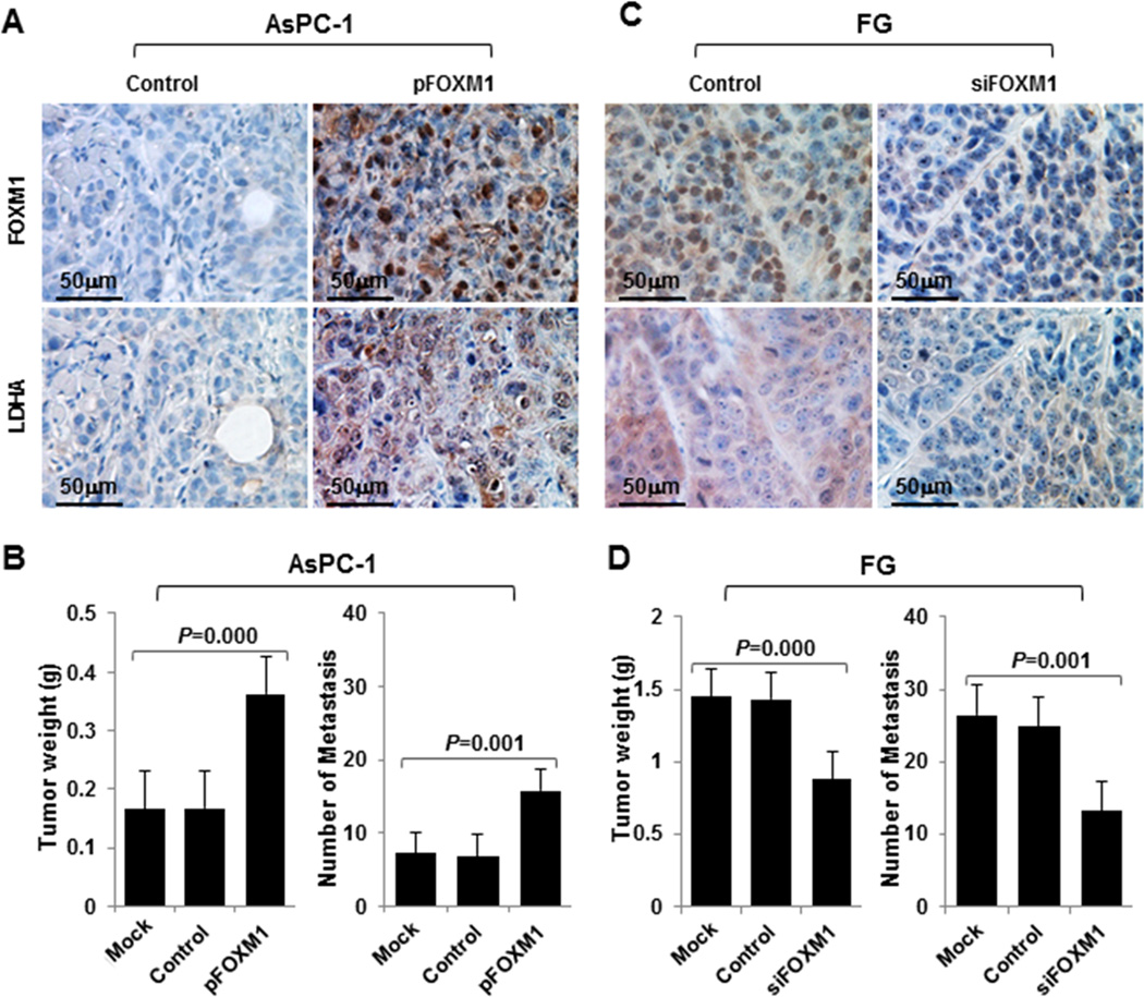
Influence of FOXM1-LDHA signaling on pancreatic cancer cell growth and metastasis in vivo. A–D, AsPC-1 cells with FOXM1 overexpression (A and B) or FG cells with knockdown of FOXM1 expression (C and D) were injected subcutaneously into the right scapular region in nude mice (1 × 10⁶/mouse, n = 5) or intravenously into the ileocolic vein in nude mice (1 × 10⁶/mouse, n = 5). The tumor-bearing mice were killed when they became moribund or on day 35 (subcutaneous) or day 21 (intravenous injection). Immunohistochemical staining of subcutaneous tumor specimens with specific anti-FOXM1 and anti-LDHA antibodies was performed. Shown are representative photos of the expression of FOXM1 and LDHA protein in AsPC-1 (A) and FG (C) cells, the weights of the tumors (B, left panel; D, left panel), and the numbers of liver surface metastases (B, right panel; D, right panel).