
Figure 6.
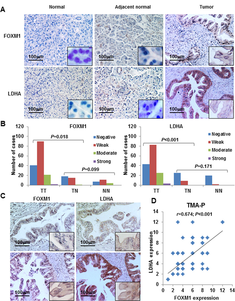
Concomitant expression of FOXM1 and LDHA in pancreatic tumors. Pancreatic tumor specimens in TMA and TMA-P were immunostained with specific anti-FOXM1 and anti-LDHA antibodies. A, representative images of FOXM1 and LDHA expression in normal pancreatic tissue, normal tumor-adjacent pancreatic tissue, and pancreatic tumor specimens (magnification, ×200). B, FOXM1 and LDHA expression in TMA-P. The expression levels were significantly higher in tumor (TT) than normal tumor-adjacent tissue (TN) and normal tissue (NN) specimens, whereas the expression of FOXM1 and LDHA did not differ between TN and NN. LDHA expression was associated with FOXM1 expression. C, representative photos of negative (upper two panels) and positive (lower two panels) FOXM1 and LDHA staining in pancreatic tumor specimens. D, direct correlation between FOXM1 expression and LDHA expression in TMA-P (n = 154; Pearson correlation coefficient, r = 0.674; P < 0.001). Some of the dots on the graph represent more than one specimen (overlapping scores).