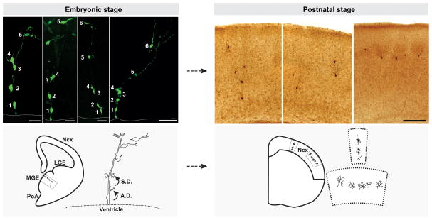
Figure 1. Clonal production and organization of neocortical interneurons.
(Left) In the embryonic MGE, RGPs divide asymmetrically at the VZ surface to self-renew and to simultaneously produce differentiating interneurons or IPs, which then divide symmetrically in the SVZ to produce differentiating interneurons. The progeny (cell 2–6) of the same RGP (cell 1) are initially closely associated with the mother RGP and organized into radially aligned clonal clusters. As development proceeds, the early-born cells progressively move away from the VZ, acquire the characteristic morphological and biophysical features of differentiating interneurons, and migrate tangentially towards the neocortex. Broken lines represent the VZ surface. (Right) After arriving at their destination in the neocortex, inhibitory interneuron clones do not randomly disperse, but form spatially organized vertical or horizontal clusters. Images of raw data are shown at the top and schematics are shown at the bottom. Scale bars (from left to right): 25 μm, 25 μm, 50 μm, 50 μm and 250 μm. Ncx, neocortex; LGE, lateral ganglionic eminence; MGE, medial ganglionic eminence; PoA, preoptic area; A.D., asymmetric division; S.D., symmetric division. Adapted from Brown et al. [20].

