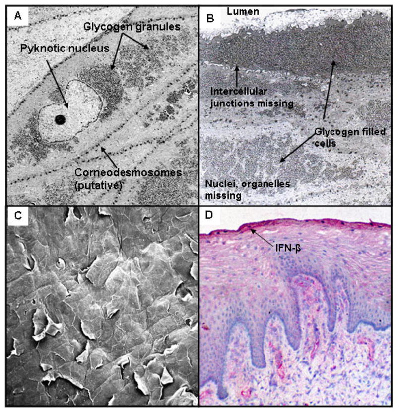
Figure 1. Comparison of morphology of epidermal vs. vaginal epithelia.

A) Structure of normal human vulvar epidermis. SB = stratum basalis; SS = stratum spinosum, SG = stratum granulosa, SC = stratum corneum.
B) Structure of normal human vaginal epithelium. (SB= stratum basalis, SuB = suprabasal layer; SC= stratum corneum).
Both specimens were collected fresh after surgery, fixed in formalin, mounted in paraffin, sectioned at 6 um and stained with hematoxylin and eosin.
