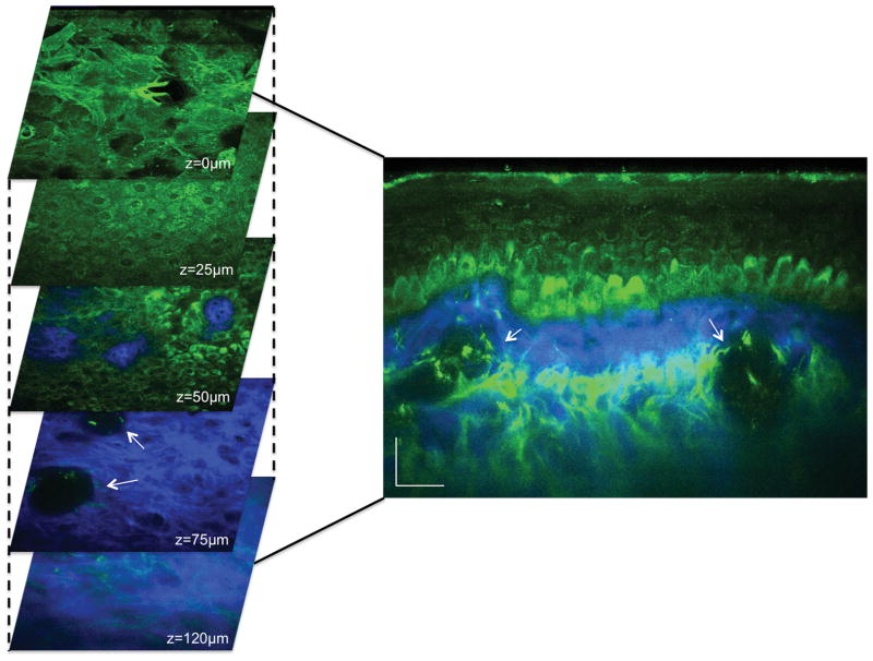
Figure 1. Pigmented normal skin.
(Left) Horizontal sections of MPM images (XY scans) at different depths showing images of the stratum corneum (z=0μm), keratinocytes normally distributed in the stratum spinosum (z=25μm), the basal cells (green) surrounding dermal papilla (blue) (z=50μm), collagen fibers (blue) and cross-sections of blood vessels (white arrows) (z=75μm), collagen (blue) and elastin (green) in the dermis (z=120μm). (Right) Cross-sectional view (XZ scan) corresponding to a vertical plane through the horizontal sections on the left. Arrows point to blood vessels.

