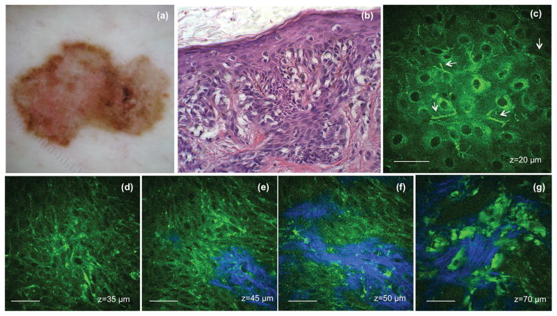
Figure 4. Melanoma - superficial spreading type.
(a) Clinical image (DermLite FOTO, Dermlite Inc.) (b) Histologic section of the lesion (c) MPM images showing ascending melanocytes (arrows) in the granulosum layer of the epidermis (d) MPM images of the basal layer showing proliferation of atypical melanocytes (highly pleomorphic melanocytes) F=0.69 (e–f) MPM images of the basal layer showing basal cells and atypical melanocytes (green) surrounding dermal papilla (blue); F=0.63 and 0.62 respectively (g) MPM images showing melanoma cells and probably melanophages invading the dermis (blue-collagen fibers). The S value for this stack is 0.35. Scale bar is 40 μm.

