
Figure 8. Comparison of the late potential and fractionated bipolar electrogram (EG) activity recorded form the epicardial (Epi) surface of the right ventricular outflow tract (RVOT) of a patient with Brugada syndrome (BrS) with similar activity recorded from coronary perfused right ventricular (RV) wedge models of BrS.
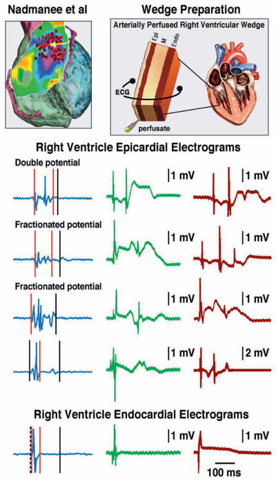
Left: Bipolar RV Epi and endocardial (Endo) EGs recorded from the RVOT of BrS patient from the study of Nademanee et al. (11). Right: Bipolar EGs recorded from the Epi surface of coronary-perfused canine RV wedge models of BrS. All recordings were obtained from preparations displaying concealed phase 2 reentry. In all cases late potentials and fractionated EG activity was the result of repolarization defects created by a inward shift in the balance currents active during the early phases of the Epi action potential. In no case did we observe primary impulse conduction delays. In both clinical and experimental models, late potentials or fractionated activity was not observed in the Endo EGs.