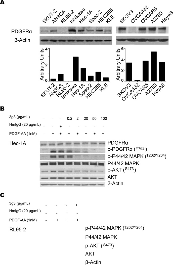
Figure 1. Expression of PDGFRa and anti-phosphorylation effect of its blockade in cancer cells.
A, Western blot analysis of PDGFRa expression in uterine and ovarian cancer cell lines. B, In vitro effect of 3G3 on PDGFRa, pPDGFRa, and its downstream targets. Cells (Hec-1A) were treated with 3G3 or nonspecific human immunoglobulin G (HmIgG) for 24 hours and then stimulated with 1 nM PDGF-AA for 10 minutes. Cell lysates were analyzed by Western blot analysis with antibodies against PDGFRa, pPDGFRa, p 44/42 MAPK, MAPK, pAKT, and AKT. C, Western blot analysis of expression of PDGFRa downstream targets in RL95-2 cells. RL95-2 cells were treated with 3G3 or PDGF-AA as mentioned above. Loading of an equal amount of sample per gel lane was confirmed by b-actin expression.

