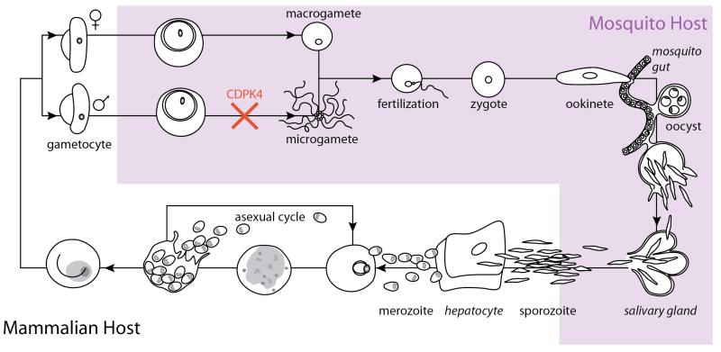
Figure 1.
Diagram of the Malaria parasite lifecycle. Asexual mammalian-host stages are shown in white and lifecycle stages that occur in the mosquito are shown in light prurple. A subset of merozoites differentiate into male and female gametocytes, which are ingested by female mosquitos upon biting an infected human. In the mosquito gut, male gametocytes divide into flagellated microgametes, which escape red blood cells (exflagellation) and swim to the macrogamete, resulting in fertilization. The resultant motile zygote forms an ookinete, which moves across the mosquito gut to form oocysts. Oocysts rupture to form sporozoites that mature in mosquito salivary glands and can be injected into humans by mosquito bites. In humans, replication via the asexual lifecycle occurs. Inhibitors of PfCDPK4, block transmission by preventing exflagellation.

