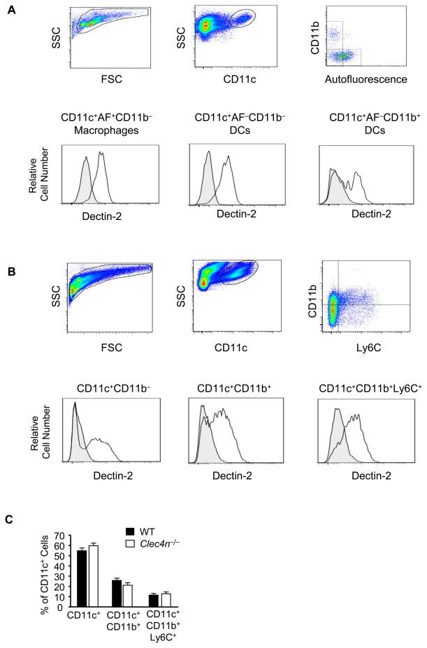
Figure 5. Dectin-2 is Specifically Expressed on CD11c+ Cells in the Lung of Naive and Df-Sensitized and Challenged Mice.
Single cell suspensions from the lung of WT C57BL/6 mice were generated and stained with the indicated antibodies or isotype controls. A. Top row shows gating of viable cells from naïve mice using FSC/SSC, staining for CD11c, and autofluorescence (AF)/staining for CD11b. Bottom row shows Dectin-2 expression (black lines) and isotype control (shaded gray) on CD11c+AF+CD11b− macrophages (left), CD11c+AF−CD11b− DCs (middle) and CD11c+AF−CD11b+ DCs (right). B. Top row shows gating of viable cells from WT C57BL/6 mice sensitized to 1 μg Df on day 0 and 1, challenged to 1 μg Df on days 14 and 15, and killed on day 17. FSC/SSC and stainings for CD11c, and CD11b/Ly6C are shown. Bottom row shows Dectin-2 expression (black lines) and isotype control (shaded gray) on CD11c+CD11b− (left), CD11c+CD11b+ (middle), and CD11c+CD11b+Ly6C+ (right) populations. C. CD11c+ subsets from WT and Clec4n−/− lung after sensitization and challenge as in B. Data are means ± SEM (n = 4-7 mice/group) combined from 2 independent experiments.

