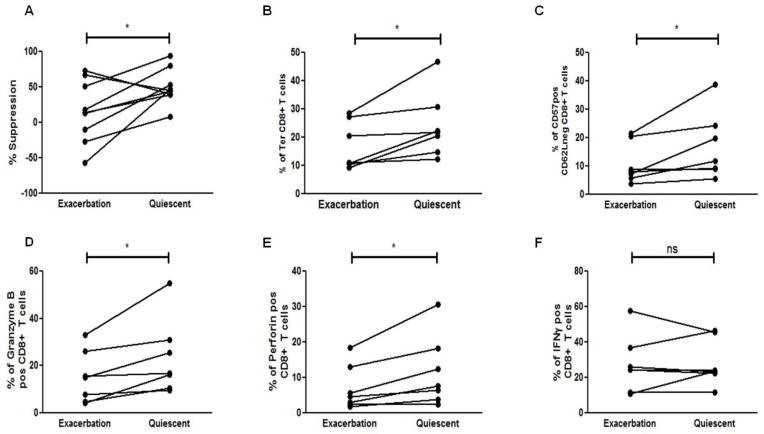
Figure 4. Phenotypic differences of neuroantigen-specific CD8+ Tregs during exacerbation vs quiescence.
(A) CD8+ T cells isolated from PBMCs of MS patients during disease exacerbation and quiescence were used in suppression assays with autologous APCs and responder CD4+CD25− T cells from disease exacerbation and quiescence respectively. (B) The frequency of terminally differentiated (Ter), CD27-CD45RO− CD8+ T cells was assessed in peripheral blood of MS during the two phases of disease and a significant increase in the percentage of Ter CD8+ T cells was observed in MS patients as they remitted. (C) MS patients showed a significant increase in the expression of CD57+CD62L− CD8+ T cells from exacerbation to quiescence. (D–F) Enriched CD8+ T cells from MS patients during either disease exacerbation or quiescence were stimulated for 5 hrs with leucocyte activation factor (LAF) (BD Pharmingen), followed by Intracellular staining for granzyme B (D), perforin (E), and IFNγ (F). A significant increase in perforin and granzyme B expression was observed in remitting MS patients while no significant change in IFNγ expression was seen (ns). *p < 0.05; PBMCs were collected from 7 RRMS patients during both disease quiescence and relapse.

