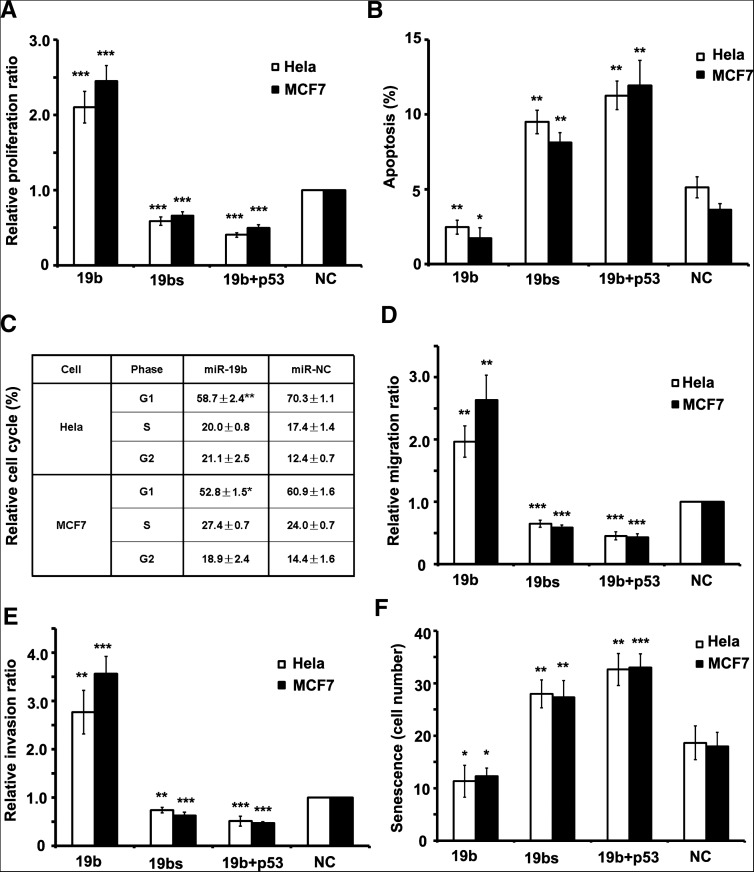
FIGURE 3.
miR-19b promotes the cell cycle, cell migration, and invasion, while inhibiting senescence and apoptosis. (A) The proliferation rate of cells as detected by EdU staining. Both Hela and MCF7 cells transfected with miR-19b divided faster than control cells, while proliferation was retarded in the sponge-transfected cells. (B) The apoptotic rate of cells was measured by Annexin-V staining. The rate of apoptosis was lower in cells transfected by miR-19b relative to the other cell types. (C) The cell cycle was detected by flow cytometry with propidium iodide (PI) staining in Hela or MCF7 cells. More cells entered G2/M phase after introducing miR-19b. Data are tested by Mann-Whitney test. (D,E) Migration (D) and invasion (E) rates detected by transwell assay showed that 19b-overexpressed cells had approximately threefold greater migration and invasion potency than NC cells, while migration and invasion rates of cells transfected with the miR-19b sponge were half as potent as NC cells. (F) Senescence rates were detected by β-galactosidase staining and the number of β-gal+ cells per microscope field were calculated. Overexpression of miR-19b reduced the rate of senescence, while miR-19b sponge drove a higher rate of senescence. In A, B, D, E, and F, all of the cell phenotypes attributed to miR-19b could be rescued by cotransfecting plasmids expressing p53 cDNA without 3′ UTR. All histograms are presented as mean ± SEM from at least three independent experiments. Data, except for C, are tested by ANOVA test and further verified by Bonferroni post-test.

