FIGURE 4.
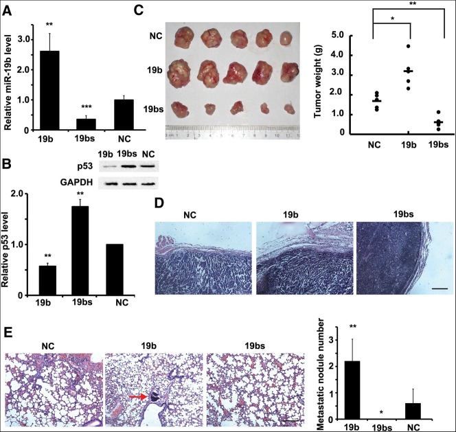
miR-19b regulates tumor growth and metastasis in vivo. (A,B) miR-19b levels (A) and p53 protein levels (B) were measured by qRT-PCR or Western blot, respectively, in three stable cell lines: Hela-19b (19b), Hela-sp (19bs), and Hela-NC (NC). (C) Three cell types were subcutaneously implanted into nude mice. n = 5. After 8 wk, mice were killed and the tumors were measured. Tumors formed by Hela-19b were the largest in size and weight, while tumors from Hela-sp cells were much smaller. (D) Haematoxylin and eosin (H&E) staining of the tumor edge. Hela-sp tumors showed a clear edge and were noninvasive. Scale bar, 50 μm. (E) H&E staining of mouse lungs, and the metastatic nodule numbers in each lung were calculated. The red arrow indicates the metastatic nodule. They were found in all of the five mice implanted with Hela-19b cells, few in those implanted with Hela-NC cells, but none in Hela-sp cells. Scale bar, 50 μm. All histograms are presented as mean ± SEM from five independent experiments. Data are tested by an ANOVA test and further verified by Bonferroni post-test.

