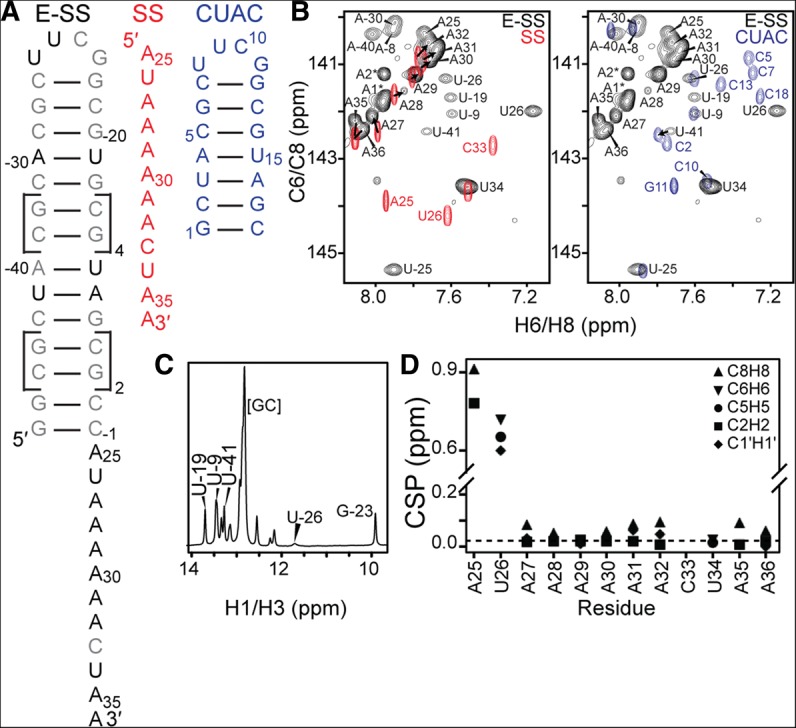
FIGURE 1.

NMR chemical shift analysis of the ssRNA–helix junction. (A) Constructs used for NMR studies and resonance assignments. (B) Overlay of 2D 1H-13C HSQC spectra of SS and E-SS allows assignment of ssRNA residues (left), while overlay of E-SS and CUAC allows assignment of helical reporter residues (right). (C) 15N-filtered 1D 1H spectrum shows characteristic imino resonances for the helix and cUUCGg tetraloop. (D) Weighted average chemical shift perturbation (CSP) of ssRNA residues between the SS and E-SS constructs shows large perturbations for ssRNA–helix junction residues, while A27–A36 have minimal perturbations.
