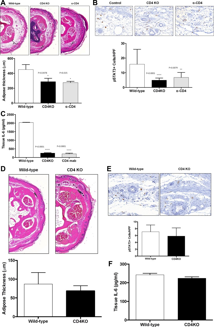
Fig. 4.
CD4+ T lymphocyte inflammation is necessary for adipose deposition and IL-6 expression. A: representative low-power (5×) cross-sectional photomicrographs and quantification of adipose tissue thickness in tails of wild-type, CD4KO, and CD4 depleted (α-CD4) mice 6 wk after tail skin/lymphatic excision. Brackets indicate subcutaneous adipose tissue thickness. B: representative high power (40×) cross-sectional photomicrographs and quantification of pSTAT3+cells/HPF in tails of wild-type, CD4KO, and α-CD4 animals 6 wk after tail skin/lymphatic excision. C: ELISA quantification of tissue IL-6 in wild-type, CD4KO, and α-CD4 tail tissues 6 wk after skin/lymphatic excision. D: representative low-power (5×) cross-sectional photomicrographs and quantification of adipose tissue thickness in tails of wild-type and CD4KO mice preoperatively. E: representative high power (40×) cross-sectional photomicrographs and quantification of pSTAT3+ cells/HPF in tails of wild-type and CD4KO mice preoperatively. F: ELISA quantification of tissue IL-6 in wild-type and CD4KO mice preoperatively.

