Fig. 5.
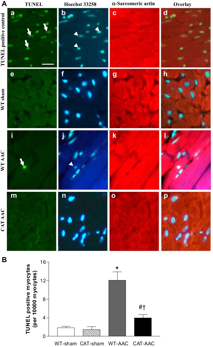
A: representative photomicrographs of LV showing terminafl deoxynucleotidyl transferase dUTP-mediated nick-end labeling (TUNEL) staining for apoptotic myocytes. Apoptotic nuclei (arrows) are shown by green fluorescence in a, e, i, and m. Nuclei (arrowheads) stained by Hoechst 33258 are shown blue in b, f, j, and n. Cardiomyocytes identified by α-sarcomeric actin staining are shown red in c, g, k, and o. The overlays in d, h, l, and p allow identification of apoptotic nuclei present in myocytes. Bar (A) = 25 μm. B: mean changes in the number of apoptotic myocytes in LV myocardium 12 wk after surgery. Values are means ± SE; n = 5–7. *P < 0.001 vs. WT-sham or CAT-sham; #P < 0.05 vs. WT-sham or CAT-sham; †P < 0.002 vs. WT-AAC.

