
Fig. 7.
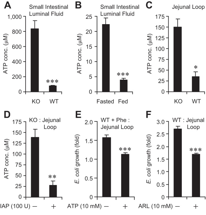
ATP inhibits bacterial growth in vivo. The small intestine was dissected out and luminal fluid was collected by gentle squeezing, then centrifuged, and the supernatant was obtained for ATP assay (see materials and methods). For determining ATP concentration and studying the effects of ATP in vivo, laparotomy was performed on mice (n = 5 per group) under general anesthesia and a 5-cm jejunal loop was constructed. Approximately 1,000 CFU of a specific bacterial species were instilled by injection into the loop. After 2 h, the loop was dissected out, homogenized, and plated on selective media for overnight growth at 37°C (see materials and methods for details). A: ATP concentrations (conc.) in the small intestinal luminal fluids of WT and IAP-KO mice. B: ATP concentrations in the small intestinal luminal fluids of WT mice fasted for 14 h. C: ATP concentrations in the jejunal loops of WT and IAP-KO mice. D: ATP concentrations in the jejunal loops of IAP-KO mice receiving IAP (100 U: injection directly into the loop). E: growth of E. coli in the jejunal loops of WT mice pretreated with 10 mM phenylalanine in the drinking water. F: growth of E. coli in the jejunal loops of WT mice receiving the ecto-ATPase inhibitor ARL 67156 (ARL; 10 mM) injection directly into the loop. Values are expressed as means ± SE. Statistics: 2-tailed Student's t-test; *P < 0.05, **P < 0.01, ***P < 0.001.