Fig. 11.
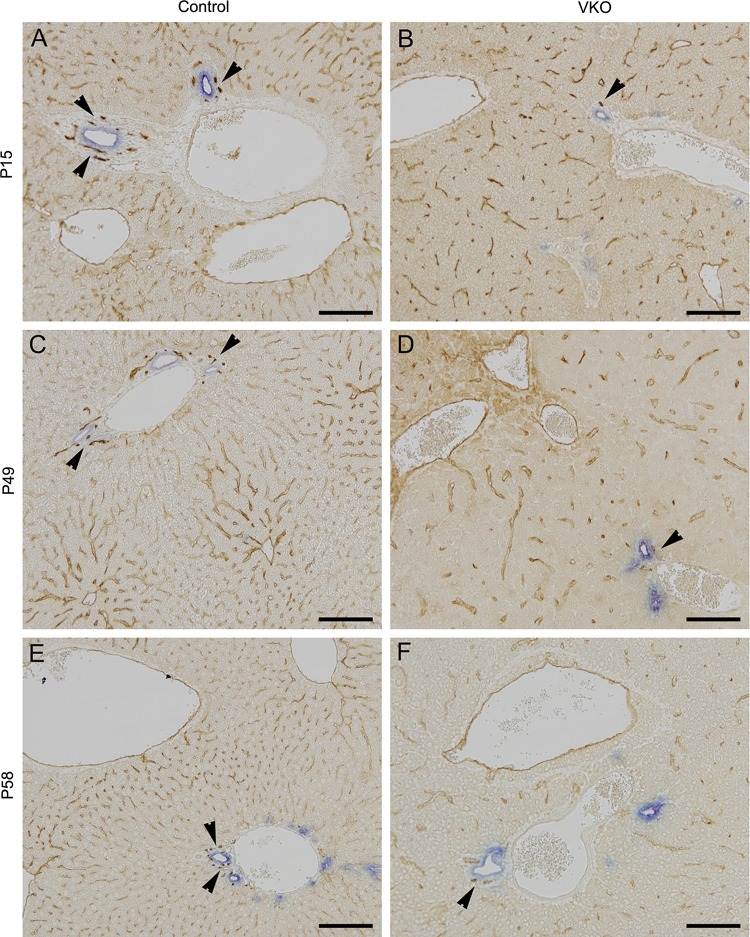
VKO mice exhibit a slight reduction in the peribiliary plexus and sinusoidal endothelial capillary network. To analyze the formation of the biliary structures and peribiliary plexus, we used cytokeratin 19 (CK19), a marker of bile ducts, and endomucin, an endothelial marker. At all stages analyzed (P15, P49, and P58), the control mice exhibit an intricate peribiliary vascular plexus (A, C, and E, arrowheads) and dense sinusoidal endothelial capillary network. In VKO mice, the peribiliary vascular plexus and sinusoidal endothelium do form. However, the peribiliary vascular plexus is simplified (B, D, and F, arrowheads), and the sinusoidal endothelial capillary network appears less dense in the VKO mice compared with littermate controls. Scale bar is 100 μm.

