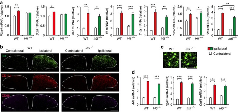
Figure 5. IRF5 is required for de novo expression of P2X4R in the spinal cord after PNI but without microglial cellular alterations.
(a) Real-time PCR analysis of mRNAs of microglial genes in the spinal cords of WT and Irf5−/− mice 7 days after PNI. Values represent the relative ratio of mRNA (normalized to the value for 18s mRNA) to the contralateral side of WT mice (n=6, *P<0.05, **P<0.01). Representative images (of three experiments) showing immunofluorescence labelling of (b) Iba1 (green), CD11b (red) and CD68 (purple) in the L4 spinal cord of Irf5−/− and WT littermates 7 days after PNI (scale bar, 200 μm), or (c) Iba1 in the L4 ipsilateral spinal cord of Irf5−/− and WT littermates 7 days after PNI (scale bar, 50 μm). (d) Real-time PCR analysis of Aif1, Itgam and Cd68 mRNA in the spinal cords of WT and Irf5−/− mice 7 days after PNI. Values represent the relative ratio of mRNA (normalized to the value for 18s mRNA) to the contralateral side of WT mice (n=6, ***P<0.001). Values are the mean±s.e.m.

