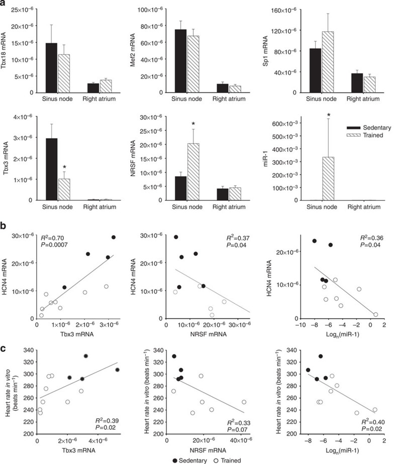
Figure 4. Three regulators of HCN4 in the sinus node are altered by training.
(a) No changes in the expression of three regulators of HCN4 in the sinus node in trained rats, but significant changes in another three. Mean (+s.e.m.) mRNA expression of Tbx18 (sinus node, n=6/6; right atrial muscle, n=7/6), Mef2 (sinus node, n=6/5; right atrial muscle, n=7/6), Sp1 (sinus node and right atrial muscle, n=6/6), Tbx3 (sinus node, n=5/8; right atrial muscle, 8/7), NRSF (sinus node and right atrial muscle, n=7/6) and miR-1 (sinus node, n=4/7; right atrial muscle, n=6/5) (normalized to expression of 18S in the case of mRNAs and RNU1A1 in the case of miR-1) in the sinus node and right atrial muscle of sedentary and trained rats shown. (b,c) Significant correlations between HCN4 mRNA (b) and heart rate in vitro (c) and Tbx3 mRNA, NRSF mRNA and miR-1 in sedentary and trained rats. Each point corresponds to a different animal. Data fit with straight lines by linear regression (n=4–8, R2 and P values shown). Student’s t-test used to test differences between data from sedentary and trained rats. Normal distribution of data was tested using the Shapiro–Wilk W-test and equal variance was tested using the F-test. When the null hypothesis of normality and/or equal variance was rejected, the non-parametric Mann–Whitney U-test was used. *P<0.05.

