Figure 1.

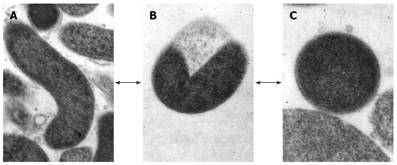
Morphological appearance of Helicobacter pylori. A: Rod-shaped; B: U-shaped; C: Coccoid form. Arrows show the hypothetical alternative pathway between the different forms. Transmission electron microscope: original magnification × 20000.

Morphological appearance of Helicobacter pylori. A: Rod-shaped; B: U-shaped; C: Coccoid form. Arrows show the hypothetical alternative pathway between the different forms. Transmission electron microscope: original magnification × 20000.