Fig. 1.
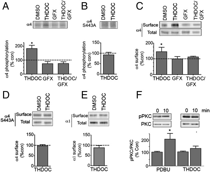
Neurosteroids regulate the phosphorylation and cell surface expression of recombinant GABAARs containing α4 subunits. (A) HEK cells expressing α4β3 receptors were labeled with 1 mCi/mL 32P-orthosphosphoric acid and treated for 10 min with DMSO (control), 100 nM THDOC, or 20 μM GFX/100 nM THDOC. Phosphorylation of α4 was measured using immunoprecipitation with subunit-specific antibodies and data normalized to vehicle-treated samples. (B) The effects of 100 nM THDOC on the phosphorylation of receptors composed of α4S443A subunits were measured as outlined above. (C) Cells expressing α4β3 subunits were treated for 10 min with DMSO (control), 100 nM THDOC, or 20 μM GFX/100 nM THDOC and labeled with NHS-biotin. The resulting cell surface and total fractions were then immunoblotted with α4 subunit antibodies. The ratio of cell surface to total α4 subunit immunoreactivity was determined and normalized to vehicle-treated control (dotted line; 100%). (D) The effects of 100 nM THDOC on the cell surface accumulation of receptors composed of α4(S443A) and β3 subunits were measured as outlined above. (E) The effects of 100 nM THDOC on the cell surface accumulation of receptors composed of α1 and β3 subunits were measured as outlined above. (F) HEK cells were treated with 100 nM PDBU or 100 nM THDOC for 10 min and then immunoblotted with pT638 and a PKC antibody that recognizes the α, βI–II, and γ subtypes of PKC. The ratio of pT638/PKC immunorecativity was determined and normalized to levels seen at t = 0. *, significantly different to control in all panels (P < 0.05; n = 4–6).

