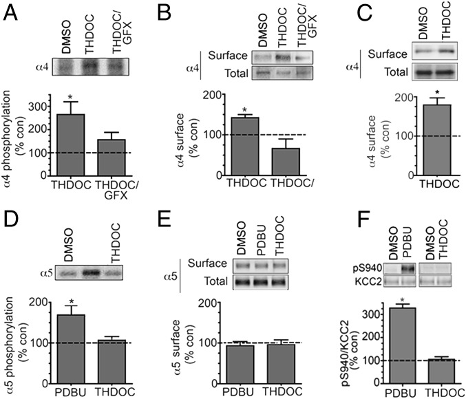
Fig. 2.
Neurosteroids selectively regulate the phosphorylation and cell surface expression of GABAARs containing α4 subunits in hippocampal slices. (A) We labeled 350 μm hippocampal slices from 8- to 12-wk-old mice with 1 mCi/mL 32P-orthosphosphoric acid and treated them for 10 min with DMSO (control), 100 nM THDOC, or 20 μM GFX/100 nM THDOC. Phosphorylation of α4 was measured using immunoprecipitation with subunit-specific antibodies and data normalized to vehicle-treated samples (dotted line; 100%). (B) Hippocampal slices were treated as above and subject to biotinylation. Cell surface and total fractions were then immunoblotted with α4 subunit antibodies. The ratio of cell surface to total α4 subunit immunoreactivity was determined and normalized to vehicle-treated controls. (C) Cell surface expression levels of the α4 subunit were determined in hippocampal slices from C57/Bl6 δ-KO mice treated for 10 min with DMSO (control) or 100 nM THDOC as detailed above. (D) Phosphorylation of the α5 subunit was measured in 32P-labeled hippocampal slices using immunoprecipitation with subunit-specific antibodies and data normalized to vehicle-treated samples. (E) Hippocampal slices were treated as above and subject to biotinylation. Cell surface and total fractions were then immunoblotted with α5 subunit antibodies. The ratio of cell surface to total α5 subunit immunoreactivity was determined and normalized to vehicle-treated controls. (F) Hippocampal slices were treated with the respective agents and then immunoblotted with pS940 and KCC2 antibodies. The ratio of pS940/KCC2 immunoreactivity was determined and normalized to vehicle-treated controls (dotted line; 100%).

