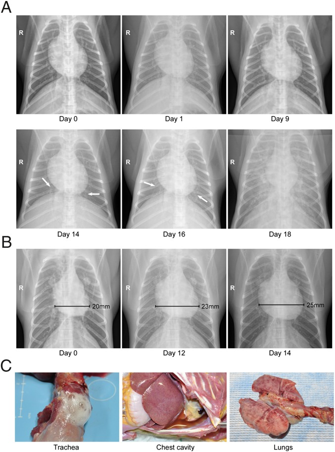
Fig. 1.
Radiographical examinations and gross pathology of macaques that developed HPS. Rhesus macaques infected with DM-SNV developed severe respiratory distress indicative of HPS. (A) Radiographic imaging. Chest X-rays were taken at regular intervals during the course of infection. Shown are serial images from a single representative animal demonstrating a rapid progression from small areas of increased density suggestive of light interstitial infiltrates to diffuse bilateral consolidation indicating severe pulmonary edema or pneumonia. (B) Right ventricle enlargement. Several animals that developed HPS demonstrated an expanded right ventricle suggestive of acute cardiac failure. (C) Gross pathology. At the time of euthanasia the majority of macaques that developed HPS presented with frothy tracheal exudates and red-tinged pleural effusions. The lungs were edematous, firm, and failed to collapse. There was multifocal consolidation and dark red discoloration that was most prevalent on the dorsal surfaces of the lung. These findings are consistent with interstitial pneumonia.

