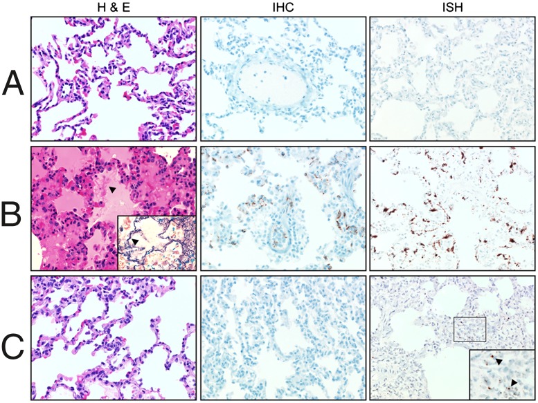
Fig. 2.
Histopathology of lungs from control and infected macaques. Rhesus macaques were inoculated with clarified lung homogenates from naive deer mice (A) or with DM-SNV (B) or VA-SNV (C). Shown are lung samples collected from representative animals in each group necropsied between 18 and 21 dpi and stained with H&E or tested for the presence of virus by IHC (anti-N antigen) or ISH (anti-N RNA). Consistent with the clinical presentation, lung samples from macaques infected with DM-SNV demonstrated multifocal to coalescing interstitial pneumonia characterized by thickening of the alveolar septae with edema, fibrin (arrowhead; Inset is stained for fibrin with phosphotungstic acid-hematoxylin), macrophages, and fewer neutrophils. Diffuse, abundant viral antigen and RNA were detected in the pulmonary endothelium in these animals. In contrast, matched samples from animals inoculated with VA-SNV demonstrated no histological abnormalities, with drastically reduced detection of viral RNA by ISH and no detectable viral antigen by IHC.

