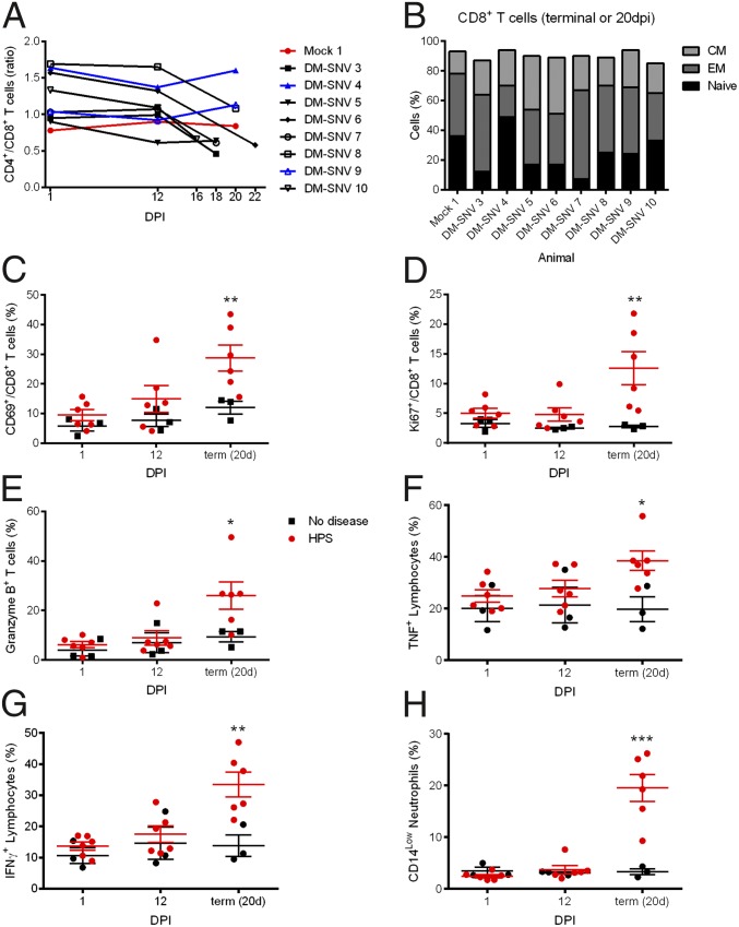
Fig. 3.
Animals that develop HPS mount a robust immune response at the time of disease. PBMCs isolated from macaques at 1, 12, and 20 dpi (for survivors) or at the time of euthanasia were stained with panels of antibodies to distinguish cell populations. (A) The ratio of CD4+ and CD8+ T cells (CD3+ cells) decreased in animals that developed HPS. (B) Memory cells were determined by their CD28 and CD95 expression on CD8+ T cells (CD3+) at the terminal stages of disease (or at 20 dpi for survivors). Animals that developed HPS had an increase in memory (effector and/or memory) cell percentages. (C–H) Cells were stained with the indicated antibodies (y axis), and percentages of cells expressing markers are shown. (E–G) PBMCs were stimulated with phorbol12-myristate13-acetate and ionomycin for 6 h, with the addition of brefeldin A for the last 4.5 h, and were stained with the indicated antibodies. Cytokine expression was increased in PBMCs from animals that developed HPS. *P < 0.05, **P < 0.01, ***P < 0.0.001.

