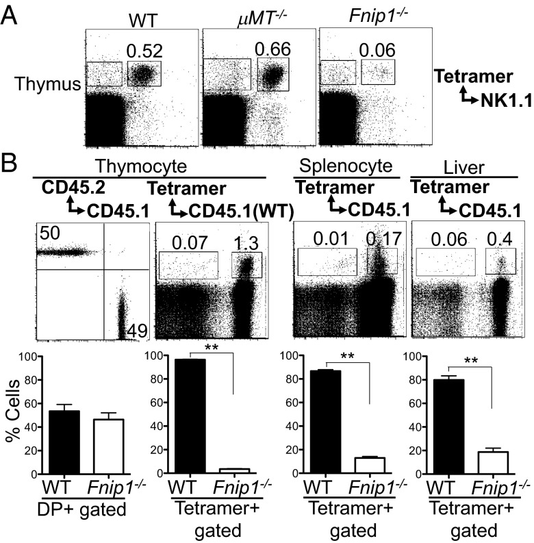
Fig. 3.
Defective iNKT cell development in Fnip1−/− mice is cell-autonomous. (A) Defective iNKT cell development in Fnip1−/− mice does not result from peripheral B-cell deficiency. Thymocytes from WT, μMT−/−, and Fnip1−/− mice were stained with antibodies specific for NK1.1 and CD1d tetramer. Shown are representative flow cytometric plots from four mice per group. (B) Bone marrow cells from Fnip1−/− (CD45.2) and WT (CD45.1) mice were mixed 1:1 and injected into irradiated Rag2−/−γc−/− recipient mice. Thymus, spleen, and liver were harvested from the recipient mice 10 wk after transplantation analyzed by flow cytometry. Shown are representative histograms (Upper) and mean ± SEM (Lower) of the percentages of cells within each gate determined from six mice per group. **P < 0.0001 (unpaired t test).

