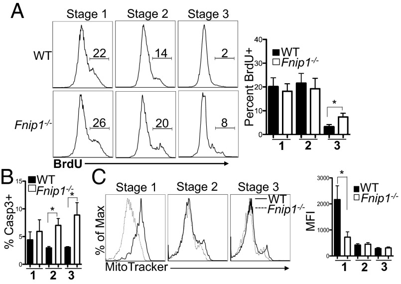
Fig. 5.
Thymic iNKT cells from Fnip1−/− mice proliferate normally but are less viable. (A) WT (Upper) and Fnip1−/− (Lower) mice were injected with BrdU and 24 h later thymocytes were harvested and tetramer-enriched for iNKT cells. Purified cells were stained with antibodies against CD44, NK1.1, and BrdU. Shown are representative flow cytometric histograms of the percentage of BrdU+ cells at each stage of iNKT cell development and the mean ± SEM from five mice per group. *P < 0.05 (unpaired t test). (B) Fnip1−/− iNKT cells have decreased viability. Thymocytes isolated from WT and Fnip1−/− mice were stained for CD44, NK1.1, CD1d tetramer, and Caspase 3. The mean ± SEM of the percentage of Caspase 3-positive stage 1, 2, and 3 iNKT cells is shown from n = 6 mice per group. *P < 0.05 (unpaired t test). (C) Fnip1−/− iNKT cells have reduced numbers of mitochondria. Thymic iNKT cells were analyzed with CD44, NK1.1, CD1d tetramer, MitoTracker green (25 nM). Mean ± SEM is shown from four mice per group. *P < 0.05 (unpaired t test).

